Fibular Hemimelia
What is Fibular Hemimelia:
- Fibular hemimelia is a congenital limb deficiency(Birth Defect).
- In this problem, part or whole of fibula bone (one of two bone present in leg, which present on outer side & thin one) is missing / not completely developed/ dysplastic.
- It is associated with hypoplasia/ dysplasia of tibia and foot.
- The phenotype has a broad spectrum of pathology varying from mild to severe limb length discrepancy, ankle/leg deformities
with or without a subtalar coalition, midfoot coalition, and absent rays. - A patient can have Limb length discrepancy, foot, and ankle deformities & deficiency, Tibial deformities, Genu Valgus & Knee instability.
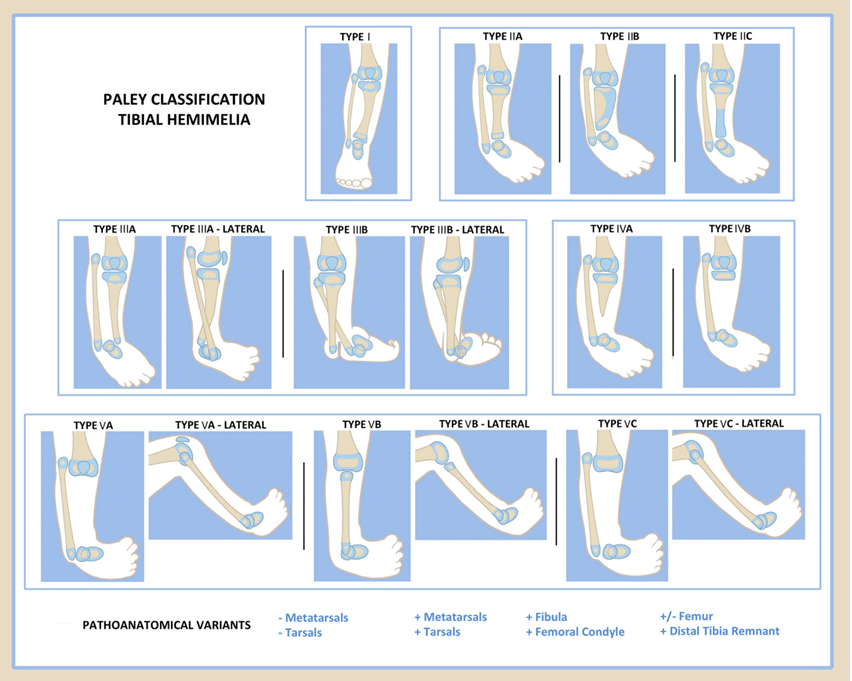
Classification of Fibular Hemimelia:
- Many classification has been given for this problem.
- Most common classification in use is given by Dr Dror Paley.
- This classification is based on patho-anatomy deformities of ankle and subtalar joint.
- Each Paley classification type has its own surgical treatment plan.
Causes of Fibular Hemimelia:
- Fibular Hemimelia present since birth.
- It is congenital.
- Most of time, causes are not known.
- Genetic factor is the most common cause for this problem.
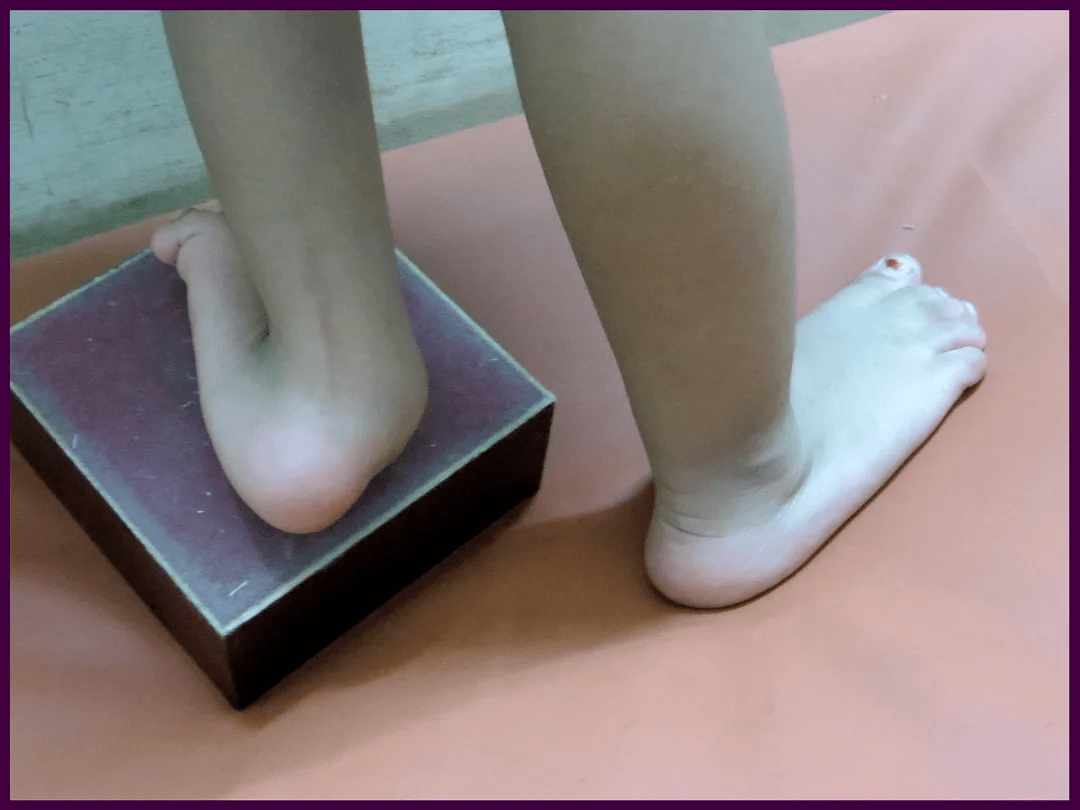
Symptoms of Fibular Hemimelia:
In fibular hemimelia, severity of fibular deficiency vary from mild shortening to complete deficiency of fibula. Child will have limping and unable to bear weight fully. So symptom vary.
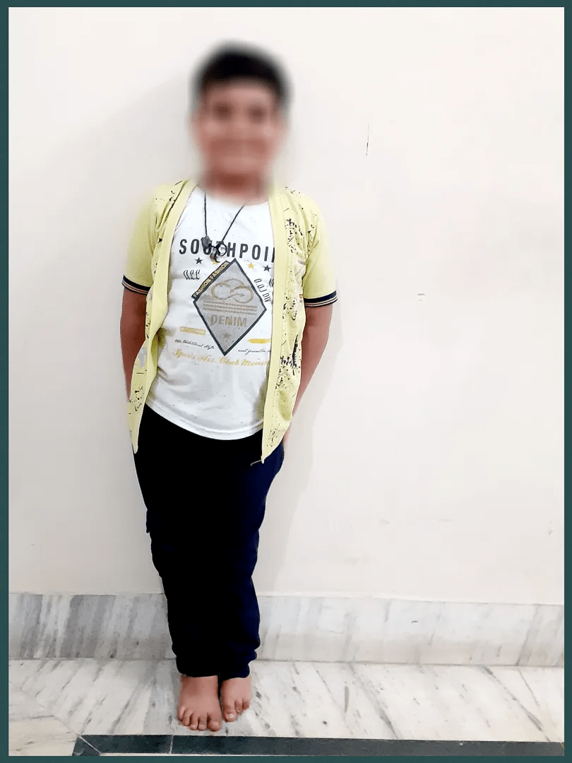
- Limb shortening
- Ankle joint deformity
- Absence of Toes (outer side) in affected foot.
- Number of missing toes can vary from one to four toes.
- Smaller size of foot.
- Displacement of heel bone to outside. On standing, double heel appearance
- Associated problem can include knee ligament deficiency & hip problem.
- Tibia can also be bend with skin puckering at anterior aspect.
How Fibular Hemimelia can be diagnosed:
- Before birth ultrasonologist can identify child with fibular hemimelia even in mother womb.
- Fibular hemimelia can be diagnosed clinically even at the time of birth.
- Confirmation of diagnosis can be made by paediatrician or paediatric orthopaedic surgeon by looking missing toes & ankle, leg deformity.
- For final documentation & confirmation of severity, X-ray of limb (leg & foot) is required.
- With the help of X-ray, We can identify severity & can also determine type of Fibular hemimelia.
- Rarely do we need MRI to see the status of foot and ankle along with missing knee ligament.
How Fibular Hemimelia can be treated:
Treatment in fibular hemimelia is depend upon severity, type & associated abnormality. If it is very mild limb length discrepancy. Then no treatment is required except reassurance and shoe raise.
In other cases, child need detail assessment. In most of the cases, child need surgical reconstruction.
Treatment need to be individualized according to problem of child. If child have deformity, trouble in standing and walking then need expert opinion of reconstructive paediatric orthopaedic surgeon. Before treatment planning, we need to look this few important aspect in child-
- Length of missing fibula
- Limb length discrepancy at maturity (check by multiplier app)
- Leg deformity
- Knee ligament status
- Ankle deformity
- Missing number of toes in foot
- Heel bone abnormality (malalignment of talus & calcaneal bone in heel)
Different Surgeries for the Treatment of Fibular Hemimelia:
Our surgical planning is based on deficiency of bony segment in foot & leg, shortening and deformity. Most of time, it is preferable to reconstruct the foot in place of amputation.
Surgeries are being done in stages.
Type of surgery is depend upon type of fibular hemimelia according to Paley classification.
Type of surgery in Fibular Hemimelia:
- Shortening osteotomy, realignment distal tibia: in this surgery tibial shortening & ankle joint alignment are being done.
- SUPER Ankle procedure (systemic utalarian procedure for extremity reconstruction): in this procedure lengthening of short tendon, excision of fibular analogue, reconstruction of foot and ankle joint are being done.
- Knee ligament reconstruction
- Toe and metatarsal osteotomy
- Limb lengthening
- Epiphysiodesis of other extremity for shortening or distal femur on same side for genu valgus deformity
Complications of Fibular Hemimelia:
- Few of the toes and finger may also be absent congenitally. If more than 3 toes are missing then child will be able to walk on our own foot.
- Ankle joint deformity can cause pain, instability and inability to run & jump.
- Limb shortening and sever deformity of foot and ankle if not managed, can cause pain, callosity & difficulty in walking.
How parents can be help?
Whenever parents see their child leg, they will be worried that what will be the future of child.
But if we consider all congenital limb deficiency, then fibular hemimelia have good prognosis for normal walking in most of the children if properly managed.
So parents need to understand their child problem and how to deal with their child.
Guideline to parent’s are-—
- Take your child to expert pediatric orthopedic surgeon in india who knows complete treatment planning.
- Discuss about treatment strategy and future outcome with your doctor
- Plan according to your feasibility, time and holiday with your busy schedule for final surgical intervention
Have an opinion about general health with your paediatrician.
For More Information: Pediatric Orthopedic Treatment FAQs
Frequently Asked Questions
What Causes Fibular Hemimelia?
In most cases, the cause of fibular hemimelia is not known. But In several studies, it is shown that this situation is related to some genetic abnormalities.
Is fibular Hemimelia a disability?
Fibular hemimelia is a defect present since birth where part or all fibular bone is not completely developed. Fibular hemimelia condition is very rare.
Will my walking be affected after surgery?
yes, but it will not produce any major problems in walking if properly done. After the surgery, you will ask to be on bed rest for few days or months. After the end of the first week, you will start therapy by the help of physiotherapists. Reconstruction require some time and even upto few months to walk

Reviewed and Submitted by Dr. Jitendra Kumar Jain
Last updated on October 30, 2020
Dr.Jitendra Jain, MD and DNB (Orthopedics), president at Trishla Foundation, an NGO for treatment of cerebral palsy, and a Consultant Pediatric Orthopedic Surgeon & Cerebral Palsy Specialist at Trishla Orthopedic Clinic & Rehab Center.
Dr. J. K. Jain is a member of the general council at Dr. SMN university of rehabilitation, Lucknow, a member of the advisory board chief commissioner for PWD, Govt. of India (New Delhi), a member of the state disability research committee (U.P.), and a member of the committee of RCI, New Delhi. He has been awarded many awards, including the Dr.Bhagawan das memorial award, the spirit of humanity award, and the state govt. award for his services towards PWD, etc. Times of India has posted his work many times and mentioned him as one of the best doctors in the field of Pediatric Orthopedics. He helped many children recovering from cerebral palsy, just like comedian jay Chanikara, who is now able to stand and walk without any support, Abena, a Ghana girl with cerebral palsy, and many more. He also organized the National Wheelchair cricket tournament and created World’s first cerebral palsy village foundation in Prayagraj. He successfully treated 10,000+ children with various kinds of orthopedic disability, conducted 160+ free assessment camps, and produced a documentary film on cerebral palsy.
Walk in Appointments Available Daily
You can make an appointment online for video tale-consultation by fixing up an appointment at this website or you can visit the clinic to make an appointment in person and show to doctor with the care of social distancing at the given time.
Contact us
Call Us
0532-3550523
+ 91 9415014994
+ 91 8577873545
+ 91 9455001645
Email Us
totrishlaortho@gmail.com
Our Location
Dr. Jitendra Kumar Jain
Trishla Orthopedic Clinic & Rehab center, 182C / 350A, Tagore Town, Prayagraj (Allahabad) U.P-211002, India