Proximal Femoral Focal Deficiency
What is Proximal Femoral Focal Deficiency?
- It is complex birth defect in which upper part of femur bone (thigh bone) is missing or malformed.
- Its severity can range from just shortening of femur to complete absence of thigh bone.
- It is very uncommon disorder and affect 1 in 20000 children.
- It is a spectrum of thigh bone deficiency, deformity, and discrepancy.
- Congenital Femoral Deficiency can have various components like Knee and hip Joint malformation, malfunction, joint instability, muscle contracture, femur bone shortening rotational deformity & cartilaginous un-ossified bone at the femur etc.
- Fibular hemimelia and Ray deficiency (absence of finger) in association with PFFD is common.
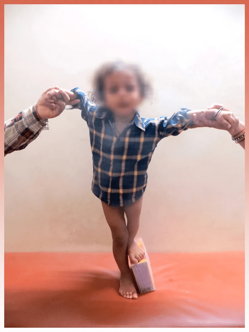
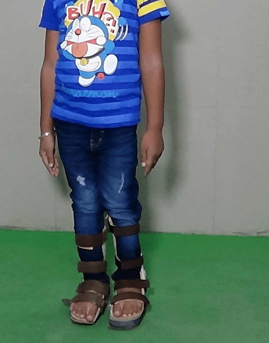
- Because of thigh bone shortening and abnormality, one limb is shorter than other.
- This difference in limb length causes most of the problem during walking.
- It is rare. Its incidence is one in two lac children.
Also Read: Unilateral Isolated Proximal Femoral Focal Deficiency
What are the causes of Proximal Femoral Focal Deficiency?
- The cause of PFFD is unknown in most cases.
- Scientist suspect that it occurs because of some insult during early phase of intrauterine life.
- This insult may occurs because of some intrauterine infection or trauma.
- Genetic association has not been proven.
- Some drug like thalidomide in the early phase of pregnancy can cause congenital limb deficiency & other birth defect.
Symptoms of Proximal Femoral Focal Deficiency:
- Significant limb shortening is the main concern. The grade of limb shoclub footrtening is dependent upon the type of PFFD.
- Hip and knee instability on same side
- The shorter length of the thigh compared to the other side,
- Not able to take weight on affected lower limb,
- A limb may be flexed at the knee and hip and rotated outside.
- Unstable hip / knee joint
- Deformity at hip & knee joint
- Associated problem like absent fibula (fibular deficiency), Plano valgus feet, club foot, congenital heart disease
- Usually it is unilateral but it may affect on both side.
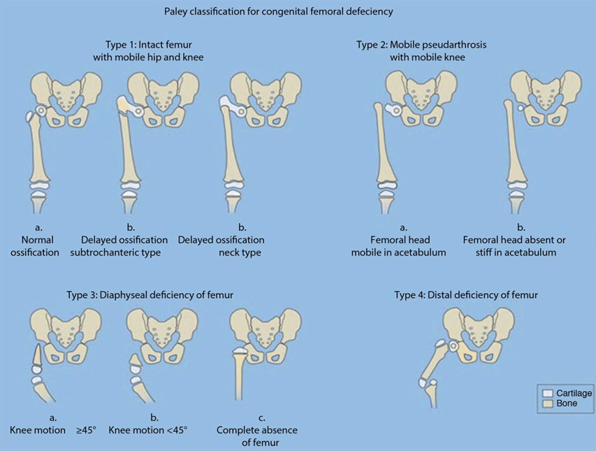
How PFFD can be Diagnosed?
- During the pregnancy, it can also be diagnosed by ultrasound in the second trimester.
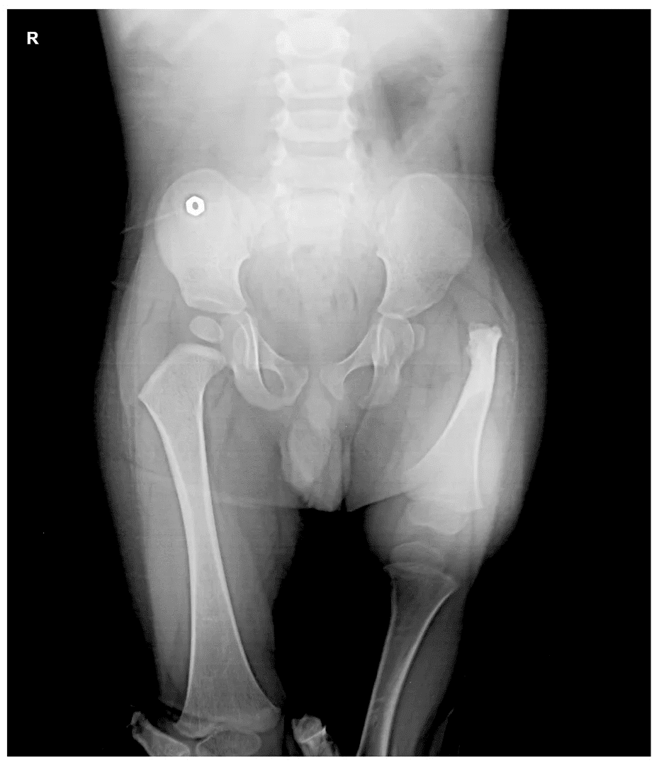
- PFFD can be diagnosed at the time of birth by observing smaller thigh length compare to other side. Affected limb will be externally rotated, flexed & abducted.
- It is very important to have detail assessment of child by the expert in the field of limb reconstruction.
- A pediatric orthopedic surgeon in India will evaluate the child fully to see the whole component of deficit and problem.
- X-ray is the initial investigation in child to define the problem.
Different tests for PFFD:
In most cases, we will need certain investigation to see the status of deficit-
X-ray-
by X-ray, we can see the bony status of the deficit. Some time some part of the femur is not so ossified so it will be very difficult to identify by X-ray.
CT scan-
CT Scan is being done to see the bony status and supplementary to MRI in elder children.
MRI-
is required to see the status of femur deficiency, a cartilaginous un-ossified portion of the femur, acetabulum joint & ligament of the knee.
Treatment for PFFD:
Treatment of PFFD is most challenging in all congenital limb deficiency disorder. It is a complex problem and present in variety of ways so every child need different set treatment planning.
Treatment are based on:
- Type of PFFD & its severity
- Stability of hip, knee & foot
- Age, overall health & associated problem
- Financial & social issues should be also be taken care during treatment planning
- Child with PFFD need multidisciplinary approach. Child need to be seen by paediatrician, paediatric orthopaedic surgeon, rehab expert & prosthetists.
- Child with sever deficiency and unrepairable problem may need prosthetic fitting for standing & walking.
- Usually treatment start when child begin to walk.
- All the reconstruction & limb lengthening will be completed in many stages before the age of maturity.
Also Read: Ensuring Surgical Safety in Proximal Femoral Focal Deficiency
Hip reconstruction:
in most of the cases with PFFD, hip joint is malformed. Proximal femur is deformed so that hip muscle are not working properly. It need detail evaluation of problem and surgery to correct proximal femur deformity, muscle balancing, improve acetabular coverage etc. This part of surgery require great expertise in this field.
Knee ligament reconstruction:
In most of the PFFD, knee ligament also absent so need ligament reconstruction. This is being done during index operation of hip reconstruction. Fascia Lata & iliotibial band (thick fascial layer under skin in lateral part of thigh) is being utilised for knee ligament reconstruction.
Treatment of associated bony anomaly:
Sometime PFFD is associated with fibular hemimelia so fibular hemimelia also need reconstruction, in which proper alignment of foot bone is being done along with excision of fibular analogue and deformity correction.
Limb Lengthening:
Before considering limb lengthening, we should be ensured many thinks like stability of hip & knee joint, predicted discrepancy of limb reconstruction. So hip knee & foot should be made align & stable by the reconstructive surgery then we need to check approximate limb length discrepancy at maturity by multiplier app. Then we can do this lengthening in 2-3 stages along with shortening of other side femur by epiphysiodesis of distal femur (temporary stoppage of growth of thigh bone on opposite side ) so that at maturity both limb will have same length. Usually first limb lengthening will be done before child goes to school (3-4 year) then at the age of 8-9 year and finally before maturity.
Prosthesis fitting:
In some cases where we are not able to align the limb due to financial, medical & social issue, then child should be given prosthetic fitting so that he can be made mobile and independent.
Reference Links :
- https://en.wikipedia.org/wiki/Proximal_femoral_focal_deficiency
- https://www.orthobullets.com/pediatrics/4043/proximal-femoral-focal-deficiency
- https://posna.org/Physician-Education/Study-Guide/Congenital-Femoral-Deficiency-(Proximal-Femoral-Fo
- https://www.researchgate.net/publication/292102267_Treatment_of_congenital_femoral_deficiency
Frequently Asked Questions
What does PFFD Means?
PFFD means Proximal focal femoral deficiency. It is a complicated birth defect in which the uppermost part of the femur bone is either malformed or missing. In this case, one leg is shorter than the other.
Different Types of Tests for PFFD?
– 1. X-Ray
- CT Scan
- MRI
4.EOS Imaging
- Ultrasound
What is the surgical procedure in a proximal femoral deficiency?
reconstruction of proximal femur and hip joint by special orthopedic surgery can be done in most of the cases

Reviewed and Submitted by Dr. Jitendra Kumar Jain
Last updated on October 30, 2020
Dr.Jitendra Jain, MD and DNB (Orthopedics), president at Trishla Foundation, an NGO for treatment of cerebral palsy, and a Consultant Pediatric Orthopedic Surgeon & Cerebral Palsy Specialist at Trishla Orthopedic Clinic & Rehab Center.
Dr. J. K. Jain is a member of the general council at Dr. SMN university of rehabilitation, Lucknow, a member of the advisory board chief commissioner for PWD, Govt. of India (New Delhi), a member of the state disability research committee (U.P.), and a member of the committee of RCI, New Delhi. He has been awarded many awards, including the Dr.Bhagawan das memorial award, the spirit of humanity award, and the state govt. award for his services towards PWD, etc. Times of India has posted his work many times and mentioned him as one of the best doctors in the field of Pediatric Orthopedics. He helped many children recovering from cerebral palsy, just like comedian jay Chanikara, who is now able to stand and walk without any support, Abena, a Ghana girl with cerebral palsy, and many more. He also organized the National Wheelchair cricket tournament and created World’s first cerebral palsy village foundation in Prayagraj. He successfully treated 10,000+ children with various kinds of orthopedic disability, conducted 160+ free assessment camps, and produced a documentary film on cerebral palsy.
Walk in Appointments Available Daily
You can make an appointment online for video tale-consultation by fixing up an appointment at this website or you can visit the clinic to make an appointment in person and show to doctor with the care of social distancing at the given time.
Contact us
Call Us
0532-3550523
+ 91 9415014994
+ 91 8577873545
+ 91 9455001645
Email Us
totrishlaortho@gmail.com
Our Location
Dr. Jitendra Kumar Jain
Trishla Orthopedic Clinic & Rehab center, 182C / 350A, Tagore Town, Prayagraj (Allahabad) U.P-211002, India